IGF-1 LR3 (Long-R3-IGF-1) is an 83-amino-acid analog of human insulin-like growth factor-1 that combines an arginine substitution at position 3 with a 13-residue N-terminal extension. These modifications reduce its affinity for IGF-binding proteins relative to native IGF-1, the structural basis researchers cite for its prolonged activity at the IGF-1 receptor in preclinical models. Apex Laboratory supplies IGF-1 LR3 strictly as a research-grade chemical reagent for in-vitro and preclinical investigation.
The defining feature of IGF-1 LR3 is an act of deliberate molecular subtraction: two engineered structural changes — an arginine substitution at position 3 and a 13-residue N-terminal extension — exist for the single purpose of weakening the molecule’s grip on the IGF-binding proteins that would otherwise hold native insulin-like growth factor-1 in check. That reduced binding is not an incidental side effect; it is the whole design. Because the IGFBP family governs the bioavailability and circulating half-life of IGF-1, and because IGFBP-3 in particular forms an acid-labile-subunit ternary complex that dramatically extends IGF-1’s plasma residence while neutralizing its acute activity,[2] an analog that escapes that binding behaves very differently in the test systems where it has been studied.
This guide examines IGF-1 LR3 as a research-grade chemical reagent: its 83-residue architecture relative to the native 70-residue peptide, the IGF-1 receptor signaling it shares with native IGF-1, the binding-protein biology that motivated its design, and the honest limits of the peer-reviewed literature that bears its name. Throughout, the framing is strictly research-context — IGF-1 LR3 is supplied for in-vitro and preclinical work only and is not a pharmaceutical, not a therapeutic, and not for human consumption.
IGF-1 LR3 at a Glance
- IGF-1 LR3 is an 83-residue analog of native 70-residue human IGF-1, built from an Arg-for-Glu substitution at position 3 plus a 13-residue N-terminal extension.
- The combined Arg3 + N-terminal-extension design markedly lowers affinity for IGF-binding proteins (IGFBPs), which is the engineered point of the molecule.
- Because IGFBPs — especially the IGFBP-3 · acid-labile-subunit ternary complex — normally sequester and clear circulating IGF-1, reduced binding is the cited basis for LR3’s prolonged action.
- Native IGF-1 in its ternary complex circulates with a greatly extended residence time, whereas free IGF-1 is cleared rapidly; LR3 shifts this balance toward a larger, longer-lived free fraction in preclinical models.
- LR3 acts at the same IGF-1 receptor tyrosine kinase as native IGF-1, engaging PI3K/Akt/mTOR and Ras/MAPK cascades; most signaling data are native-IGF-1-derived, not LR3-specific.
- IGF-1 LR3 is research-use-only and is a different molecule from native-sequence recombinant human IGF-1 (mecasermin/Increlex); it is not for human or veterinary use.
IGF-1 LR3
What Is IGF-1 LR3? Definition and Structural Overview
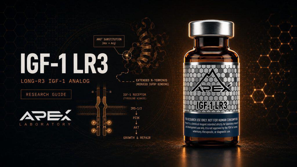
IGF-1 LR3 — written in the literature as Long-R3-IGF-1 or LR3IGF-I — is an 83-amino-acid recombinant analog of human insulin-like growth factor-1. Native human IGF-1 is a 70-residue single-chain polypeptide; the LR3 analog is built on that backbone but carries two engineered changes. The first is an arginine-for-glutamate substitution at position 3 (the “R3” of the name). The second is a 13-residue N-terminal extension — Met-Phe-Pro-Ala-Met-Pro-Leu-Ser-Ser-Leu-Phe-Val-Asn — appended ahead of the native sequence (the “Long”). The arithmetic is straightforward: 70 native residues plus the 13-residue extension yields the 83-residue analog, consistent with the catalog molecular weight of roughly 9,117.5 g/mol.
The 83-residue Long-R3 analog architecture
The two modifications work in concert. Neither the position-3 change nor the N-terminal extension alters the core IGF-1 fold that engages the receptor; instead, both reshape the surfaces that the IGF-binding proteins recognize. The net structural consequence, repeatedly described in the in-vivo literature, is an analog “which has much reduced affinities for IGF binding proteins,” as Conlon and colleagues at the University of Adelaide put it in their guinea-pig infusion study.[6]
Specifications and an honest caveat on sourcing
The molecular specifications above — CAS 946870-92-4, molecular weight near 9.1 kDa, and the residue sequence — are catalog-derived and cross-checked for internal consistency rather than independently confirmable through a public small-molecule registry; IGF-1 LR3 is a large protein analog and does not resolve to a PubChem CID. The molecular formula is therefore listed as “Not specified” rather than asserted. Researchers should treat the lot-specific Certificate of Analysis as the authoritative identity record, a point developed in the quality-verification section below and in our guide to reading a peptide COA. For broader context on where this reagent sits, see the growth-hormone-axis research peptides hub.
IGF-1R Receptor Tyrosine Kinase and Downstream Signaling
Whatever modifications IGF-1 LR3 carries on its periphery, the receptor it engages is the same one native IGF-1 uses: the type 1 insulin-like growth factor receptor (IGF-1R), a transmembrane receptor tyrosine kinase. Ligand binding to the extracellular domains triggers a conformational change that drives autophosphorylation of the intracellular kinase domains, creating docking sites that recruit adaptor proteins and launch two principal intracellular cascades.
IGF-1R receptor tyrosine kinase → PI3K/Akt/mTOR and Ras/MAPK (ERK)
IGF-1 LR3 engages the type 1 IGF receptor (IGF-1R), a receptor tyrosine kinase. Ligand binding drives receptor autophosphorylation and recruitment of adaptor proteins, branching into two cascades: PI3K/Akt/mTOR, which promotes protein synthesis and — via Akt-mediated inhibition of FoxO transcription factors — suppresses E3-ubiquitin-ligase-driven proteolysis; and Ras/MAPK (ERK), associated with proliferative programs. These maps derive chiefly from native IGF-1 studies; LR3 is understood to act at the same receptor.[1]
The PI3K/Akt/mTOR pathway
The first cascade runs through phosphoinositide 3-kinase (PI3K), Akt, and the mechanistic target of rapamycin (mTOR). In a 2020 review of IGF-1 in skeletal muscle, Yoshida and Delafontaine describe how IGF-1 increases protein synthesis through PI3K/Akt/mTOR and PI3K/Akt/GSK3-beta signaling, and how the same Akt activity inhibits FoxO transcription factors to suppress the E3 ubiquitin ligases that drive proteolysis.[1] This anabolic/anti-catabolic balance is why IGF-1 signaling is so heavily studied in muscle-biology research models.
The Ras/MAPK (ERK) pathway
The second cascade proceeds through the Ras/mitogen-activated protein kinase (MAPK) axis, terminating in ERK activation, and is associated in the published literature with proliferative and mitogenic cellular programs. It is worth stating plainly: these pathway maps are derived overwhelmingly from native IGF-1 studies.[1] IGF-1 LR3 is understood to act at the same receptor and engage the same downstream machinery, but LR3-specific signaling characterization is comparatively limited, and this guide does not present native-IGF-1 findings as direct LR3 evidence.
IGF-Binding Protein (IGFBP) Biology and Bioavailability
To understand why IGF-1 LR3 was engineered the way it was, the binding-protein layer has to come first. In the circulation, IGF-1 does not travel freely. Baxter’s foundational 1994 review describes a family of six IGF-binding proteins (IGFBP-1 through IGFBP-6) that regulate the bioavailability and half-life of the IGFs.[2]
The IGFBP-3 / ALS ternary complex
The dominant carrier is IGFBP-3, which combines with the acid-labile subunit (ALS) to form a high-molecular-weight ternary complex. In that complex, as Baxter details, the hypoglycemic potential of the bound IGF is neutralized and its circulating half-life is greatly extended.[2] The ternary complex is, in effect, a circulating reservoir: it keeps a large pool of IGF-1 in the bloodstream but holds it in a relatively inactive, slowly-released form.
How IGFBPs govern clearance and the free fraction
The quantitative link between binding-protein status and pharmacokinetics has been shown directly in humans. Mauras and colleagues, studying recombinant human IGF-1 pharmacokinetics in hypopituitarism, found that clearance and volume of distribution correlate reciprocally with IGFBP-3 and ALS levels — underscoring the critical role of binding proteins in modulating IGF-1 bioavailability in vivo.[4] In other words, the binding proteins are not passive passengers; they are the principal determinant of how long IGF-1 persists and how much of it is free to act. That insight is the lever the LR3 design pulls.
LR3 Analog Design Rationale: Engineering Reduced IGFBP Binding
If IGFBPs govern clearance and the free fraction, then an analog that binds them poorly should, in principle, present a larger and longer-lived pool of receptor-available growth factor. This is the explicit design logic behind the IGFBP-resistant variant class, and it has been tested directly through protein engineering.
Why Arg3 and the N-terminal extension lower IGFBP affinity
Genentech’s Dubaquié and colleagues engineered binding-protein-selective IGF-1 variants and showed that variants with reduced IGFBP affinity exhibit altered biodistribution and clearance as a direct function of that affinity.[5] Their work establishes the central principle in clean form: change how tightly an IGF-1 molecule is held by its binding proteins, and you change where it goes and how fast it is cleared. The Arg3 substitution and N-terminal extension of LR3 are two structural means to that same end.
The IGFBP-resistant variant class
IGF-1 LR3 is not an isolated invention but one member of a family of IGFBP-resistant variants that includes des(1-3)IGF-I, R3-IGF-I, and Long-IGF-I. Tomas and colleagues at the CRC for Tissue Growth and Repair in Adelaide explicitly named long-R3IGF-I (LR3IGF-I) among the variants that bind poorly to IGF-binding proteins.[3] Treating the analogs as a class is methodologically important, because several of the load-bearing pharmacokinetic observations come from the sister analog des(1-3)IGF-I rather than from LR3 itself — a distinction this guide preserves rather than blurs. The relationship between IGF-1 LR3 and the related mechano-growth-factor splice variant is explored further in our IGF-1 LR3 and PEG-MGF comparison guide.
Half-Life of IGF-1 LR3 vs Native IGF-1 (Explicit Comparison)
Because this is the question researchers ask most often, it deserves a direct, carefully hedged answer. The short version: native IGF-1’s effective duration is dominated by its binding-protein status, and IGF-1 LR3 was engineered specifically to escape that constraint — but the precise numbers are far better established for native IGF-1 than for LR3.
IGFBP binding as the primary half-life determinant
For native IGF-1, the binding-protein layer sets two very different clocks. Free, unbound IGF-1 is cleared rapidly — on the order of minutes — while IGF-1 sequestered in the IGFBP-3 / ALS ternary complex persists far longer, with a greatly extended circulating half-life.[2] Mauras and colleagues quantified the in-vivo coupling, showing that recombinant human IGF-1 clearance tracks reciprocally with IGFBP-3 and ALS.[4] The practical reading is that native IGF-1’s longevity is borrowed from its carriers.
What is observed for LR3 — and what is not
For the IGFBP-resistant variants, Tomas and colleagues reported that analogs binding poorly to IGFBPs — long-R3IGF-I among them — showed roughly two-to-threefold greater potency and four-to-eightfold more prolonged action than native IGF-1 in pigs and marmoset monkeys, framed specifically around hypoglycemic action.[3] That is the strongest directly-comparative evidence for LR3’s prolonged activity, and it is preclinical. Critically, an exact human half-life value for IGF-1 LR3 is not well established in the peer-reviewed literature; the “longer half-life” claim should be read as a preclinical, comparative observation rooted in reduced IGFBP binding, not as a clean human pharmacokinetic constant. Researchers planning reconstitution and handling should consult our peptide reconstitution guide and peptide storage guide rather than infer stability from circulating half-life figures.
IGF-1 LR3 vs Native IGF-1 vs Mecasermin (Native rhIGF-1)
| Attribute | IGF-1 LR3 (Long-R3) | Native human IGF-1 | Mecasermin (Increlex) |
|---|---|---|---|
| Sequence length | 83 residues (analog) | 70 residues | 70 residues (native sequence) |
| Key modifications | Arg3 substitution + 13-residue N-terminal extension | None (reference) | None (recombinant native rhIGF-1) |
| IGFBP affinity | Markedly reduced (engineered) | High (forms IGFBP-3/ALS ternary complex) | Native (high, IGFBP-bound) |
| Reported duration of action | Prolonged vs native in preclinical models (e.g., 4-8x for IGFBP-resistant variants, hypoglycemic action) | Short free half-life; extended in ternary complex | Native rhIGF-1 PK |
| Regulatory category | Research-use-only reagent; no approved indication | Endogenous peptide / research context | FDA & EMA-approved biologic (severe primary IGF-1 deficiency) |
Preclinical Research Findings With LR3 and IGFBP-Resistant Analogs
The directly-on-target LR3 literature is concentrated in a handful of 1990s in-vivo infusion studies, several from the Adelaide group, and they are most valuable for what they reveal about context-dependence rather than for any simple narrative of uniform potency.
In-vivo infusion studies across species
Conlon and colleagues infused long-R3IGF-I in guinea pigs and observed that it stimulated organ growth while paradoxically reducing plasma IGF-I, IGF-II, and IGF-binding-protein concentrations — consistent with an analog that bypasses the normal binding-protein buffering.[6] Steeb and colleagues compared IGF-I peptides, including the IGFBP-resistant analog, for trophic effects on the small-intestinal epithelium in rats, documenting tissue-specific proliferative signaling in a preclinical model.[9]
The species-dependence caveat
The most important honesty check comes from Dunaiski and colleagues, who reported that analogs binding poorly to IGFBPs stimulate growth in rats but inhibit growth in pigs, while long-R3IGF-I suppressed plasma growth hormone, IGFBP-3, and endogenous IGF-I in pigs.[7] This directional reversal across species is a direct warning against over-generalizing LR3’s effects as uniformly anabolic. A complementary line of evidence comes from the sister analog: Sun and colleagues studied the biodistribution of radiolabeled des(1-3)IGF-I in tumor-bearing mice and found its tissue distribution and blood clearance governed by IGFBP-3 complex formation, reinforcing the general principle that reduced IGFBP binding reshapes biodistribution.[8] Note that this is des(1-3)IGF-I work cited for the analog-class rationale, not as direct LR3 data.
IGF-1 Signaling in Skeletal Muscle: Hypertrophy and Satellite Cells
Much of the research interest in IGF-1 LR3 traces to the well-characterized role of native IGF-1 in skeletal-muscle biology. This section summarizes that native-IGF-1 physiology, on the understanding that LR3 acts on the same axis but lacks dedicated LR3-specific muscle trials.
IGF-1/Akt/mTOR and protein synthesis
As Yoshida and Delafontaine detail, IGF-1 drives muscle protein synthesis through PI3K/Akt/mTOR signaling while simultaneously restraining proteolysis by inhibiting FoxO-mediated transcription of muscle-specific E3 ubiquitin ligases.[1] The dual action — building protein while suppressing its degradation — is the mechanistic reason IGF-1 sits at the center of so many hypertrophy and atrophy research models.
Satellite cell activation and plasticity
Skeletal-muscle plasticity also depends on satellite cells, the resident muscle stem cells. Snijders and colleagues reviewed satellite-cell biology in human muscle, describing their role in repair, remodeling, and adaptation.[12] Moore and colleagues subsequently associated dysregulated IGF-1 expression with a blunted satellite-cell response after exercise with age, linking the IGF-1 axis to satellite-cell dynamics in human physiology.[13] Both are native-IGF-1 studies; they establish the biological backdrop against which IGFBP-resistant analogs are investigated, not direct evidence of LR3 activity. For upstream comparators that act on the same axis from a different angle, see our Ipamorelin research guide, Sermorelin research guide, and CJC-1295 research guide.
Distinction From Native rhIGF-1 and Mecasermin (Increlex)
A recurring point of confusion deserves a clear, unambiguous answer: IGF-1 LR3 is not the same molecule as the FDA-approved IGF-1 biologic, and it is not a formulation of it. This is the inverse of a same-molecule precedent — the approved product is a structurally different protein.
Mecasermin is native-sequence recombinant human IGF-1
Mecasermin, marketed as Increlex, is recombinant human IGF-I in its native 70-residue sequence. Keating’s 2008 review confirms mecasermin as recombinant human IGF-I approved in the United States and the European Union for severe primary IGF-I deficiency.[10] Kemp’s 2009 review similarly situates rhIGF-1 (mecasermin) clinical use in children with growth-hormone insensitivity and IGF-1 deficiency.[11]
Why IGF-1 LR3 is a different molecule, not a formulation
IGF-1 LR3, by contrast, carries the Arg3 substitution and the 13-residue N-terminal extension that make it an 83-residue analog with deliberately reduced IGFBP affinity. Because the two share neither sequence nor regulatory category, no same-molecule pharmaceutical comparison applies here: mecasermin is an approved pharmaceutical with a defined indication, while IGF-1 LR3 is a research-use-only reagent with no approved therapeutic indication in any jurisdiction. Apex Laboratory positions IGF-1 LR3 strictly within the research-grade reagent category; for the general principle of how research-grade chemicals differ from approved pharmaceuticals, see our editorial standards.
Dosing and Handling Considerations in Research Settings
This section covers reagent handling only. It provides no human or veterinary dosing guidance of any kind; IGF-1 LR3 is for in-vitro and preclinical research exclusively.
Reconstitution and storage of lyophilized powder
IGF-1 LR3 is supplied as a lyophilized powder. General research-handling practice for the lyophilized material is storage at −20°C, with the reconstituted solution kept at 2–8°C for short-term use and at −20°C for longer-term aliquots, while avoiding repeated freeze–thaw cycles that can degrade peptide integrity. These are general peptide-handling parameters drawn from catalog specifications rather than compound-specific published stability studies, and researchers should follow their own validated protocols. Step-by-step procedure is covered in our reconstitution guide and storage guide.
In-vitro versus in-vivo preclinical context
In published research, IGFBP-resistant analogs have been used both as cell-culture supplements — where the reduced IGFBP binding can change effective free-ligand exposure in serum-containing media — and in animal infusion models such as those described by Conlon and Dunaiski.[6][7] The species-dependence documented in those studies is a reminder that experimental design choices materially affect outcomes and should be reported transparently.
Quality Verification: COA, HPLC Purity, and ESI-MS Identity
For any IGF-1 analog, reproducible research begins with confident identity and purity, because a mischaracterized or impure reagent quietly corrupts every downstream result.
Why ≥99% HPLC purity matters for reproducibility
Reversed-phase high-performance liquid chromatography (HPLC) resolves the target analog from truncation products, deletion sequences, and other process-related impurities, and a stated purity threshold — commonly reported at greater than or equal to 99 percent — quantifies how clean the preparation is. The methodology behind this metric is covered in our HPLC purity testing guide.
ESI-MS mass confirmation of the 83-residue analog
Purity alone does not establish that the molecule is the one intended. Electrospray-ionization mass spectrometry (ESI-MS) confirms molecular identity by matching the measured mass against the expected mass of the 83-residue analog — near 9.1 kDa — distinguishing IGF-1 LR3 from native IGF-1 or other variants that might co-elute. Our mass-spectrometry verification guide details how this identity check is read on a certificate of analysis. These quality metrics speak to reagent identity and lot-to-lot consistency only; they make no statement about any therapeutic property.
Research Limitations and Open Questions
Sound research framing requires naming what the literature does and does not support. For IGF-1 LR3 specifically, several limitations are worth stating plainly.
Thin LR3-specific literature and reliance on extrapolation
The directly-on-target LR3 literature is genuinely thin and largely preclinical, anchored on 1990s animal-infusion studies from the Adelaide CRC for Tissue Growth and Repair.[3][6][7] Much of the mechanistic picture — IGF-1R signaling, PI3K/Akt/mTOR, satellite-cell biology, and IGFBP regulation — is necessarily anchored on native IGF-1 and analog-class work,[1][2][5] and should not be read as LR3-specific evidence.
Outstanding pharmacokinetic and specification uncertainties
Exact human half-life figures for IGF-1 LR3 are not established in the peer-reviewed record; the prolonged-activity claim is comparative and preclinical. The species-dependence shown by Dunaiski — growth stimulation in rats versus inhibition in pigs[7] — cautions against treating LR3 as uniformly anabolic. Finally, the catalog molecular weight and CAS number could not be independently confirmed through a public small-molecule registry, and the molecular formula is left unspecified rather than asserted. These are open questions, and a transparent research program treats them as such. Further reading is collected in our research library.
Safety, Tolerability & Adverse-Event Observations (Research Context)
Because IGF-1 LR3 is a research-use-only reagent with no approved human or veterinary indication, there is no clinical safety dossier for the analog itself. What the published record does contain are adverse-event and tolerability observations from preclinical animal infusion studies of long-R3IGF-I and from the broader biology of reduced IGFBP binding. These are reported below strictly as research findings — not as patient side-effects, not as a guide to what any subject would experience, and not as advice of any kind.
Hypoglycemia as the dominant documented tolerability signal
In published animal research, the most consistently characterized acute effect of IGFBP-resistant IGF-1 variants is hypoglycemic action. Baxter’s circulation review explains the mechanistic basis: native IGF-1 carries intrinsic hypoglycemic potential that is normally neutralized when the peptide is held in the IGFBP-3 / acid-labile-subunit ternary complex.[2] An analog engineered to escape that buffering therefore presents more free, insulin-receptor-cross-reactive growth factor. Tomas and colleagues quantified the consequence directly, reporting that variants binding poorly to IGFBPs — long-R3IGF-I among them — produced roughly two-to-threefold more potent and four-to-eightfold more prolonged hypoglycemic action than native IGF-1 in pigs and marmoset monkeys.[3] In a research setting, this is the single most important documented tolerability characteristic of the analog class.
IGF-axis suppression and species-dependent effects in animal studies
Animal infusion work also documents effects on the endogenous growth-hormone/IGF-1 axis. Conlon and colleagues observed that long-R3IGF-I infusion in guinea pigs reduced circulating plasma IGF-I, IGF-II, and IGF-binding-protein concentrations even as it stimulated organ growth — a feedback pattern consistent with an analog that bypasses normal binding-protein buffering.[6] Dunaiski and colleagues reported the same suppressive direction in pigs, where long-R3IGF-I lowered plasma growth hormone, IGFBP-3, and endogenous IGF-I, and — critically — inhibited rather than stimulated growth in that species despite stimulating growth in rats.[7] This species-directional reversal is itself a tolerability caveat: the analog’s net effect is model-dependent, and preclinical observations in one species do not transfer cleanly to another. No fabricated adverse-event rates, no human safety claims, and no therapeutic conclusions can be drawn from these studies; they are reported here only to characterize what the peer-reviewed literature documents.
Sourcing Research-Grade IGF-1 LR3
Reagent quality is upstream of every conclusion a study can draw, which is why sourcing belongs in any serious discussion of IGF-1 LR3. Apex Laboratory supplies IGF-1 LR3 as a lyophilized chemical reagent intended for in-vitro and preclinical research only.
The purity and identity trust angle
Each lot is characterized to a target of ≥99% purity verified by reversed-phase HPLC, with molecular identity confirmed by electrospray-ionization mass spectrometry (ESI-MS) against the expected mass of the 83-residue analog. Per-batch results are documented on a Certificate of Analysis available through the lab-verified COA archive, and our broader editorial and quality posture is described in our editorial standards. We do not publish placeholder lot numbers or fabricated analysis dates; the COA that ships with a given lot is its authoritative record. For the adjacent reagents most often studied alongside IGF-1 LR3 — the PEGylated mechano-growth-factor variant and the upstream secretagogue Ipamorelin — see the comparison resources linked throughout this guide and the products below.
IGF-1 LR3 and Adjacent Research Reagents
IGF-1 LR3
Long-R3 IGF-1 analog (Arg3 substitution + 13-residue N-terminal extension) with reduced IGFBP binding; supplied at ≥99% purity verified by HPLC and mass spectrometry.
View ProductPEG-MGF
PEGylated mechano-growth-factor splice variant of IGF-1; muscle and tissue-recovery research reagent.
View ProductIpamorelin
Selective growth hormone secretagogue; upstream GH/IGF-1 axis research comparator.
View ProductFrequently Asked Questions
What is IGF-1 LR3?
IGF-1 LR3, or Long-R3-IGF-1, is an 83-amino-acid analog of native human insulin-like growth factor-1. It carries an arginine substitution at position 3 and a 13-residue N-terminal extension, modifications reported to lower its affinity for IGF-binding proteins. It is supplied as a research-grade chemical reagent for in-vitro and preclinical investigation only, not for human or veterinary use.
How is IGF-1 LR3 different from native IGF-1?
Native human IGF-1 is a 70-residue peptide. IGF-1 LR3 adds a 13-residue N-terminal extension and an Arg-for-Glu substitution at position 3, producing an 83-residue analog. In published preclinical work, these changes reduce binding to IGF-binding proteins (IGFBPs). Because IGFBPs normally sequester circulating IGF-1, this structural difference is the basis for the analog’s reported altered pharmacokinetic behavior in research models.
What is the half-life of IGF-1 LR3 and why is it longer than native IGF-1?
The reported longer activity of IGF-1 LR3 relative to native IGF-1 is generally attributed to its reduced affinity for IGF-binding proteins. Because IGFBPs sequester and clear free IGF-1, an analog that binds them poorly can show more prolonged action, as observed in preclinical infusion studies. Exact human half-life values are not well established in the peer-reviewed literature, and most data come from animal models of related IGFBP-resistant variants.
How is research-grade IGF-1 LR3 reconstituted and stored?
In research settings, lyophilized IGF-1 LR3 is typically reconstituted with a suitable sterile diluent and gently dissolved without vigorous agitation, following the researcher’s own validated protocol. General handling practice stores the lyophilized powder at minus 20 degrees Celsius, keeps the reconstituted solution at 2 to 8 degrees Celsius for short-term use or minus 20 degrees Celsius for longer-term aliquots, and avoids repeated freeze-thaw cycles. These are general peptide-handling parameters, not compound-specific stability data.
What adverse effects have been documented for IGF-1 LR3 in research?
There is no human safety dossier for IGF-1 LR3. In published animal studies, the most consistently documented effect of IGFBP-resistant IGF-1 variants is hypoglycemic action, reported as more potent and prolonged than native IGF-1. Infusion studies also recorded suppression of endogenous growth hormone, IGFBP-3, and IGF-1, with effects that differed by species. These are preclinical research observations only, not patient side-effects or any indication of human safety.
How does IGF-1 LR3 signal at the cellular level?
Like native IGF-1, IGF-1 LR3 is understood to act at the IGF-1 receptor, a receptor tyrosine kinase. Ligand binding triggers receptor autophosphorylation and activates the PI3K/Akt/mTOR and Ras/MAPK (ERK) cascades in published native-IGF-1 studies. These pathways regulate cellular processes such as protein synthesis and survival. Note that most signaling data are derived from native IGF-1; LR3-specific mechanistic studies are comparatively limited.
Is IGF-1 LR3 the same as mecasermin (Increlex)?
No. Mecasermin (Increlex) is native-sequence recombinant human IGF-1, an FDA-approved biologic. IGF-1 LR3 is a structurally distinct 83-residue analog with an N-terminal extension and Arg3 substitution. They are different molecules in different regulatory categories: mecasermin is an approved pharmaceutical, while IGF-1 LR3 is a research-use-only reagent with no approved therapeutic indication in any jurisdiction.
How is the identity and purity of research-grade IGF-1 LR3 verified?
Research-grade IGF-1 LR3 is typically characterized by reversed-phase HPLC to confirm purity (commonly reported at greater than or equal to 99 percent) and by electrospray-ionization mass spectrometry (ESI-MS) to confirm the molecular mass of the 83-residue analog. A certificate of analysis documenting these results supports experimental reproducibility. These quality metrics speak to reagent identity and consistency only, not to any therapeutic property.
Continue Your Research
Researchers building broader context across the Apex Research Library may find the following references useful:
- Growth Hormone Axis Research Peptides — The cluster hub situating IGF-1 LR3 among GH/IGF-1 axis reagents.
- IGF-1 LR3 vs PEG-MGF — Lateral comparison of the Long-R3 analog with the PEGylated mechano-growth-factor variant.
- Ipamorelin Research Guide — Upstream selective GH secretagogue studied as a GH/IGF-1 axis comparator.
- Sermorelin Research Guide — GHRH(1-29) analog acting upstream of hepatic IGF-1 production.
- CJC-1295 Research Guide — Long-acting GHRH analog for axis-level mechanism context.
- How to Read a Peptide COA — Interpreting the certificate of analysis that documents identity and purity.
- HPLC Purity Testing — How reversed-phase HPLC quantifies peptide purity for reproducibility.
- Mass Spectrometry Verification — How ESI-MS confirms the molecular identity of the 83-residue analog.
Research Use Disclaimer
IGF-1 LR3 is supplied by Apex Laboratory strictly as a research-grade chemical reagent for in-vitro and preclinical laboratory investigation only. It is not a drug, not a dietary supplement, and not a pharmaceutical product; it is not for human or veterinary use, diagnostic use, or consumption of any kind. Nothing in this article constitutes medical advice, a therapeutic claim, or a representation that IGF-1 LR3 is safe or effective for any use in humans or animals. IGF-1 LR3 is a structurally distinct analog and is not the same molecule as native-sequence recombinant human IGF-1 (mecasermin/Increlex), which is a separately regulated, approved biologic; the two should not be conflated. All cited findings are research-context observations, predominantly from preclinical and native-IGF-1 studies, and do not establish any clinical effect. Researchers are responsible for compliance with all applicable institutional, local, and national regulations governing the handling of research chemicals.