Mass spectrometry verifies peptide identity by ionizing the molecule into the gas phase, separating ions by mass-to-charge ratio (m/z), and matching the observed mass against the theoretical target. Common ionization methods are electrospray (ESI) and matrix-assisted laser desorption (MALDI); analyzer classes include quadrupole, time-of-flight, Orbitrap, and QTOF. Combined with HPLC, MS confirms ≥99% identity in research-grade material.
Mass spectrometry confirms peptide identity through one of the cleanest cascades in analytical chemistry. A peptide enters as solute and leaves as data: ionization ejects the molecule into the gas phase as a charged ion (electrospray sprays charged droplets that desolvate; MALDI vaporizes a matrix-embedded sample with a laser pulse). The mass analyzer then sorts ions by mass-to-charge ratio — a quadrupole filters, a time-of-flight tube measures drift time, an Orbitrap records oscillation frequency. The detector counts ions, the instrument plots intensity against m/z, and the resulting peak position resolves to a molecular mass that either matches the theoretical mass calculated from the peptide’s sequence or doesn’t. Match means identity confirmed. Mismatch means something else is in the vial.
This article walks the cascade. Mass spectrometry is the orthogonal-to-HPLC verification methodology that confirms what molecule occupies the main HPLC peak; Apex Laboratory’s MS verification standard, paired with HPLC for ≥99% purity, appears as a worked example of the methodology rather than the article’s selling point. The criteria are vendor-agnostic; the chemistry organizes the article.
Peptide Mass Spectrometry at a Glance
- Mass spectrometry confirms peptide identity through a discrete-step cascade — ionization to gas-phase ions, mass analyzer separation by m/z, detection, spectrum, and theoretical-vs-observed mass match — every step has a defined physical principle and the final spectrum is a single-shot identity signature
- The mass-to-charge ratio (m/z) is the central measurement; charge state is determined from isotope spacing on the spectrum (1.0 Da spacing = +1 charge; 0.5 Da = +2; 0.33 Da = +3), and charge-state assignment is a prerequisite for resolving molecular mass from the observed m/z value
- Electrospray ionization (ESI) is preferred for sequence confirmation via tandem MS/MS through LC-coupled multiply-charged ions; MALDI is preferred for rapid mass screening through MALDI-plate singly-charged single-shot workflows; both produce intact peptide ions for identity confirmation
- Mass analyzer classes span quadrupole (~1,000-3,000 resolution) for targeted screening, time-of-flight (~10,000-40,000) for MALDI-coupled identity workflows, Orbitrap (~100,000-500,000) for modern proteomics, and quadrupole-TOF hybrids for tandem MS/MS sequence verification
- Tandem MS/MS provides sequence-level confirmation through precursor ion selection, collision-induced dissociation, b-ion / y-ion fragment series, and a sequence walk via consecutive mass differences; database matching engines (Mascot, Sequest) score candidate matches probabilistically
- HPLC measures purity %, MS confirms identity of the main peak — the two methods answer different questions, and the canonical research-grade ≥99% verification standard pairs them as orthogonal methodologies
What Mass Spectrometry Measures: The m/z Principle
Mass spectrometry is the analytical methodology that measures the mass-to-charge ratio (m/z) of ions in the gas phase. For a peptide, the experiment produces an ion of the intact molecule (typically a protonated species [M+H]+, [M+2H]2+, or [M+nH]n+ depending on charge state) and measures the m/z of that ion through the mass analyzer’s physical separation principle. The Aebersold and Mann 2003 Nature review codified MS as the dominant peptide-identification methodology in modern proteomics, and the methodology has only deepened since.1
The m/z value resolves to molecular mass once charge state is known: m × z = mass observed at that m/z. A peptide with a calculated monoisotopic mass of, say, 1,500 Da appears as [M+H]+ at m/z 1,501.0078, as [M+2H]2+ at m/z 751.0078, and as [M+3H]3+ at m/z 501.0078 — three different m/z observations of the same molecule at three different charge states. The Mann, Hendrickson, and Pandey 2001 Annual Review of Biochemistry tutorial review codifies the m/z-to-mass arithmetic alongside the underlying mass-analyzer physics at a level that has become canonical for the proteomics methodological literature.5
The single experimental output is a spectrum: m/z on the x-axis, ion intensity on the y-axis, with peaks at the m/z values where ions populated the analyzer. The spectrum’s peak positions resolve to molecular masses; the molecular masses resolve to molecular identities; identity confirmation is the final cascade output.
Why MS for Peptide Identity Verification
Mass spectrometry verifies peptide identity by measuring molecular mass and matching the observed value against the theoretical mass calculated from the intended peptide’s sequence. The intended peptide produces a known [M+H]+ or [M+nH]n+ ion; the observed mass either matches within instrument tolerance or it doesn’t. Match means the molecule in the vial is the molecule the sequence specified.1
This is a categorically different verification question from HPLC purity. HPLC measures peak-area distribution — what fraction of the chromatogram is the main peak. MS measures molecular mass — what molecule the main peak actually is. A ≥99% HPLC peak whose mass does not match the intended peptide is a high-purity sample of the wrong molecule. The Anicetti and Hancock 1994 Bioprocess Technology review of analytical considerations in protein purification establishes the analytical-philosophy framing for the orthogonal-verification logic that has persisted into the contemporary research-grade peptide standard.9
MS also detects mass-shift impurities that HPLC may miss. Methionine oxidation produces a +16 Da shift (Met to methionine sulfoxide) or +32 Da (Met to methionine sulfone) and may co-elute with the main HPLC peak depending on column conditions. Asparagine deamidation produces a +1 Da shift (Asn to Asp/iso-Asp) that is often below HPLC resolution under standard mobile-phase conditions. Deletion sequences produce mass shifts equal to the missing residue’s mass and may co-elute with the parent peptide if the deletion is internal and the chromatographic separation is incomplete.5
The orthogonal-method framing is canonical: HPLC quantifies purity, MS confirms identity, and the contemporary ≥99% research-grade peptide standard requires both methods to be applied per batch. The depth-treatment of HPLC sits in the Understanding HPLC Testing for Peptide Purity guide; the depth-treatment of the ≥99% standard sits in the same-batch sister article Why ≥99% Purity Matters.
Ionization Methods: ESI vs MALDI
The first stage of every peptide MS experiment is ionization — converting the dissolved or matrix-embedded peptide into a gas-phase ion the mass analyzer can sort. Two soft-ionization methods dominate contemporary peptide MS: electrospray ionization (ESI) and matrix-assisted laser desorption ionization (MALDI). Both produce intact peptide ions suitable for identity confirmation; the choice depends on whether sequence-level verification or rapid mass screening is the question.
Electrospray ionization (ESI) sprays peptide solution through a fine needle held at high voltage, producing charged droplets that desolvate as they traverse a heated capillary into the mass analyzer. ESI is a soft ionization method — peptide bonds remain intact during the gas-phase transition — and it produces multiply-charged ions [M+H]+, [M+2H]2+, [M+3H]3+ for larger peptides. The multiply-charged-ion behavior is a feature: charge state is inferred from isotope spacing in the spectrum, and multiply-charged ions enter the mass-analyzer’s resolvable m/z range even for peptides too large to detect at +1 charge.5 ESI is LC-coupled — the standard proteomics workflow is LC-MS/MS, where reverse-phase HPLC separates peptides upstream of the ESI source, and tandem MS/MS sequence confirmation runs downstream.1
Matrix-assisted laser desorption ionization (MALDI) vaporizes a matrix-embedded sample with a UV laser pulse; the matrix absorbs the laser energy and transfers protons to the peptide, producing predominantly singly-charged [M+H]+ ions. MALDI is also soft, and its singly-charged-ion behavior produces simpler spectra than ESI for many peptides. MALDI is plate-coupled — samples are spotted onto a MALDI plate with co-crystallized matrix, and a high-throughput laser-rastering workflow can analyze hundreds of spots in an hour.2
ESI is preferred for sequence confirmation via tandem MS/MS through its LC-coupled multiply-charged-ion workflow that fragments cleanly and supports modern Orbitrap-class resolution. MALDI is preferred for rapid mass screening through its singly-charged simple spectra and high-throughput plate workflow. Both produce intact peptide ions for identity confirmation; the choice depends on the question.
Mass Analyzer Types: Quadrupole, TOF, Orbitrap, QTOF
The second stage of the cascade is the mass analyzer — the physical device that sorts ions by mass-to-charge ratio. Four analyzer classes appear with meaningful frequency across contemporary peptide MS: quadrupole, time-of-flight (TOF), Orbitrap, and quadrupole-TOF (QTOF) hybrids. The Domon and Aebersold 2006 Science review covers the instrumentation classes and their application to peptide and protein analysis at a level that remains canonical in the analytical-chemistry methodology literature.3
Quadrupole analyzers use four parallel rod electrodes with oscillating RF and DC voltages to filter ions by m/z — only ions of a selected m/z reach the detector at any given moment, and scanning the voltages walks through m/z values to build the spectrum. Quadrupoles are robust, fast, and produce modest resolution (~1,000-3,000 resolving power); they are the workhorse front-end for targeted peptide MS and for the precursor-selection stage of tandem MS/MS hybrids.
Time-of-flight (TOF) analyzers accelerate ions through a fixed potential and measure the time required to traverse a drift tube of known length. Time of flight is proportional to the square root of m/z; TOFs achieve resolution of ~10,000-40,000 with modern reflectron geometries. MALDI-TOF is the canonical high-throughput peptide identity instrument.
Orbitrap analyzers trap ions in an electrostatic field around a central spindle electrode; ions oscillate axially at frequencies proportional to the inverse square root of m/z, and the oscillation is detected as image current. Orbitrap-class resolution reaches ~100,000-500,000 — the modern proteomics ultra-high-resolution standard. The Hardman and Makarov 2003 Analytical Chemistry paper interfaced the Orbitrap analyzer to an electrospray ion source, making the analyzer practical for LC-MS/MS peptide workflows.7
Quadrupole-TOF (QTOF) hybrids combine a quadrupole front-end (for precursor ion selection) with a TOF back-end (for fragment-ion analysis) — the canonical configuration for tandem MS/MS sequence verification. The 2016 update to Aebersold and Mann’s MS-proteomics review documents the modern instrumentation landscape where QTOF and Orbitrap-class hybrids dominate sequence-level peptide identification.2
Five Peptide MS Method Configurations
| Method | Ionization | Resolution | Best Use |
|---|---|---|---|
| ESI-Quadrupole | Electrospray ionization; multiply-charged ions; LC-coupled | ~1,000-3,000 resolving power; nominal-mass accuracy | Targeted peptide screening; quantitative SRM/MRM workflows; precursor-selection front-end for tandem MS hybrids |
| ESI-TOF | Electrospray ionization; multiply-charged ions; LC-coupled | ~10,000-40,000; accurate-mass capability via reflectron geometry | LC-MS workflows requiring accurate mass without ultra-high resolution; mid-throughput peptide identity confirmation |
| ESI-Orbitrap | Electrospray ionization; multiply-charged ions; LC-coupled | ~100,000-500,000; ultra-high mass accuracy (typically ≤5 ppm) | Modern proteomics ultra-high-resolution peptide identity and quantification; mass-shift impurity discrimination at high accuracy |
| MALDI-TOF | Matrix-assisted laser desorption ionization; predominantly singly-charged ions; plate-coupled | ~10,000-40,000; accurate-mass capability via reflectron | High-throughput peptide identity screening; rapid mass-fingerprinting workflows; large peptides where multiply-charged ESI distributions complicate analysis |
| QTOF (hybrid) | Electrospray ionization; multiply-charged precursor; LC-coupled | ~10,000-40,000 (TOF stage); accurate-mass fragments | Tandem MS/MS sequence verification; b-ion / y-ion fragment analysis; database-matched peptide sequencing |
Reading a Peptide MS Spectrum
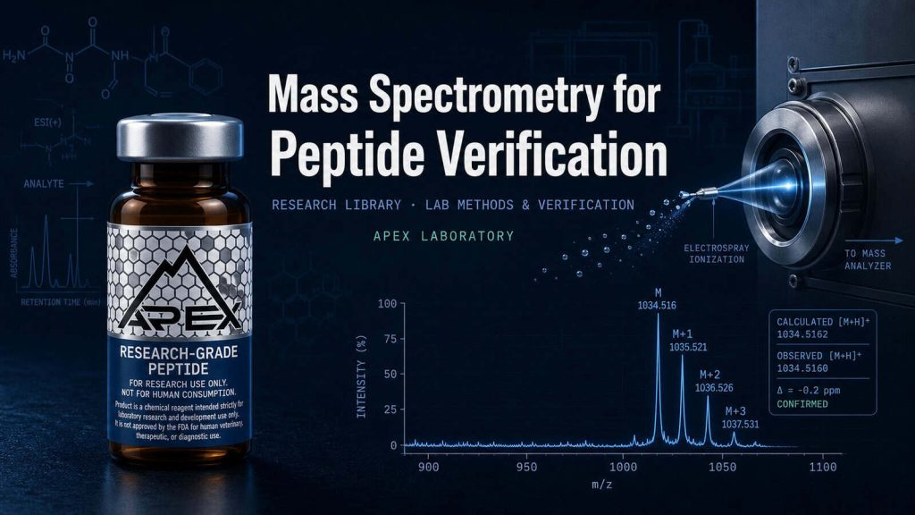
The peptide MS spectrum is a two-axis plot: m/z on the x-axis, relative intensity on the y-axis (typically normalized to the base peak at 100%). The reader’s task is to identify the dominant charge state, the monoisotopic peak, the isotope envelope, and the calculated-versus-observed mass match.
Dominant charge state depends on peptide size. Small peptides (<2,500 Da) typically appear as [M+H]+ singly-charged species; medium peptides (2,500-5,000 Da) as [M+2H]2+; larger peptides at +3, +4, or higher charge states.5 ESI typically produces a distribution of charge states; the dominant peak is selected for mass calculation.
Monoisotopic peak is the lowest-mass peak in the isotope envelope — corresponding to the molecule constructed from the most abundant isotope of each element (12C, 1H, 14N, 16O, 32S). Higher-mass peaks at M+1, M+2, M+3 correspond to species incorporating one, two, or three 13C / 15N / 2H natural-abundance heavy isotopes.
Isotope spacing resolves charge state directly. Spacing between consecutive isotope peaks equals 1/z Da: spacing of 1.0 Da indicates +1 charge; 0.5 Da indicates +2; 0.33 Da indicates +3; 0.25 Da indicates +4. The Steen and Mann 2004 Nature Reviews Molecular Cell Biology tutorial codifies the isotope-spacing-to-charge-state inference alongside the broader peptide-sequencing literacy that the article anchors.4
Theoretical mass calculation sums the monoisotopic residue masses, adds 18.0106 Da for terminal water, and adds 1.00784 × z Da for protonation at charge state z. The calculated value is compared against the observed monoisotopic peak’s m/z (multiplied by z); match within instrument tolerance (e.g., ±5 ppm for Orbitrap, ±100 ppm for low-resolution quadrupole) confirms identity.
Tandem MS/MS for Peptide Sequence Verification
Tandem mass spectrometry — MS/MS — extends the methodology from molecular-mass measurement to peptide-sequence determination. The single MS measurement confirms what the molecule weighs; tandem MS confirms what amino acid residues, in what order, the molecule is built from. The Steen and Mann 2004 Nature Reviews Molecular Cell Biology tutorial review on peptide sequencing remains the canonical pedagogical reference for the methodology and its ion nomenclature.4
Step 1 — Precursor ion selection. The first mass analyzer (typically a quadrupole or the first stage of a QTOF) selects the peptide ion of interest from the source — the “precursor” — by isolating it at its m/z value and excluding other ions in the source from the next stage.
Step 2 — Collision-induced dissociation (CID) or higher-energy collisional dissociation (HCD). The selected precursor enters a collision cell where it collides with neutral gas (typically nitrogen or argon). Collisions deposit internal energy that causes peptide-bond cleavage along the backbone. The Krishnamurthy, Hunt, and Yates 1989 PNAS paper on tandem MS structural characterization of cyclic peptides documents the historical lineage in which Hunt and Yates established tandem MS/MS as a peptide-sequencing methodology in the late 1980s.8
Step 3 — Fragment ion series (b-ions and y-ions). Backbone cleavage produces two complementary ion series. B-ions retain the charge on the N-terminal fragment, numbered from the N-terminus (b1, b2, b3, etc.). Y-ions retain the charge on the C-terminal fragment, numbered from the C-terminus (y1, y2, y3, etc.). Each consecutive b-ion (or y-ion) differs from the previous by the mass of one amino acid residue.
Step 4 — Sequence walk via mass differences. The difference in mass between consecutive b-ions (or y-ions) equals the residue mass of the amino acid at that position. Walking the b-ion or y-ion series from low to high m/z reads out the peptide sequence directly from the spectrum.
Step 5 — Database matching or de novo identification. The observed fragment-ion pattern is matched against theoretical patterns calculated from a sequence database, or sequenced de novo from mass differences without database reference. Mascot and Sequest are the canonical database-search engines; Mascot’s probability-based scoring approach was codified in the Perkins, Pappin, Creasy, and Cottrell 1999 Electrophoresis paper that established the algorithmic framework.6
MS in the Verification Workflow with HPLC
The most counterintuitive feature of contemporary peptide verification is how naturally HPLC and MS pair as orthogonal methods. They answer different questions and resolve different impurity classes; the combination gives the full picture for ≥99% research-grade verification.1
HPLC measures purity %. The reverse-phase chromatogram separates the main peptide from synthesis impurities; peak integration computes main-peak area divided by total peak area. The number is a verifiable claim about the analytical method’s resolution of the chromatographic peak distribution. The Hancock-lineage analytical-philosophy literature establishes the framing — a purity number is a statement about the method, not an absolute property of the material.9
MS confirms identity of the main peak. The molecular mass measured for the eluting main peak should match the theoretical mass calculated from the intended peptide’s sequence. Match within instrument tolerance answers the orthogonal verification question — is the main peak the right molecule.
MS detects HPLC-coelution impurities. Diastereomers from epimerization may co-elute with the parent peptide; MS distinguishes them via different fragmentation patterns even when masses match. Methionine oxidation (+16 Da) may co-elute under certain mobile-phase conditions; MS resolves the +16 signal cleanly. Deletion sequences with shifted retention may not separate from the main peak on HPLC; MS reveals via the expected-mass mismatch.
The two together establish ≥99% by HPLC + MS-confirmed identity as the canonical research-grade verification standard. The depth-treatment of HPLC’s role sits in the Understanding HPLC Testing for Peptide Purity guide; the depth-treatment of the ≥99% threshold sits in the same-batch sister article Why ≥99% Purity Matters.
What Mass Spectrometry Doesn’t Tell You
MS is a powerful methodology, and like every analytical method it has scope limitations. Educational discipline requires naming them honestly.
Purity quantification limits. MS ionization efficiency varies by molecule; relative ion abundance does not reliably reflect relative concentration in a mixture. A trace impurity that ionizes efficiently can produce a more intense peak than a major component that ionizes poorly. Purity quantification is HPLC’s job, not MS’s.2
Ionization suppression in mixtures. Co-eluting species can suppress ionization of the analyte under ESI conditions; absence of evidence in MS is not evidence of absence in solution. The matched-pair LC-MS workflow mitigates this by separating species before ionization.
Identity confirmation tolerance. Mass match is within instrument tolerance — typically ±5 ppm for Orbitrap, ±20-50 ppm for QTOF, ±100 ppm for low-resolution quadrupole. Rare isobaric species that share nominal mass but differ in elemental composition can match at low resolution and resolve only at high resolution.
Stereochemistry blindness. D-amino acid and L-amino acid have identical molecular mass; MS does not distinguish stereoisomers. Diastereomers from racemization require specialized chiral chromatography upstream, or MS/MS fragmentation pattern analysis where the diastereomers fragment differently.4
MS in Research-Grade Peptide QC: Apex Educational Framing
The reader who has worked through the cascade now has a chemistry-anchored framework for evaluating any research-peptide vendor’s MS verification claim. The framework is vendor-agnostic; it applies to Apex Laboratory and to every other compliant vendor in the research-grade reagent category equally.10
A research-grade peptide COA should reference MS verification specifically, not just “MS confirmed.” The discipline-aligned verification trace identifies the ionization method (ESI or MALDI), the analyzer class (quadrupole, TOF, Orbitrap, QTOF), the calculated theoretical mass, the observed mass, and the tolerance window of the match (e.g., ±5 ppm). The depth-treatment of COA reading sits in the How to Read a Peptide Certificate of Analysis guide; vendor MS verification claims that omit method specificity should be evaluated against the same percentage-stated, instrument-named, per-batch discipline that the analytical-philosophy literature has codified for HPLC.9
Apex Laboratory’s verification standard pairs HPLC ≥99% with orthogonal MS identity confirmation on every batch, with per-lot certificate of analysis documenting the verification. Apex’s MS verification appears in this article as one factual illustration of what research-grade MS verification looks like under the methodology described above; the framework would equally validate any compliant vendor operating to the same standard. The same-batch sister articles complete the cluster: How to Evaluate a Peptide Vendor operationalizes the verification questions; Research-Grade vs Pharmaceutical-Grade Peptides covers the regulatory-category distinction; Why ≥99% Purity Matters covers the purity-threshold rationale paired with this article’s MS-methodology depth.
Research-Grade Catalog Examples Under HPLC + MS Verification
BPC-157
Apex Laboratory BPC-157 is supplied at ≥99% purity, with HPLC and orthogonal mass spectrometry verification on every batch and per-lot certificate of analysis. For in-vitro and preclinical research only — not for human consumption.
View Product →Semaglutide
Apex Laboratory research-grade Semaglutide is supplied at ≥99% purity, with HPLC and orthogonal mass spectrometry verification on every batch and per-lot certificate of analysis. Distinct from approved pharmaceutical formulations; the Apex catalog product is for in-vitro and preclinical research only — not for human consumption.
View Product →Retatrutide
Apex Laboratory research-grade Retatrutide is supplied at ≥99% purity, with HPLC and orthogonal mass spectrometry verification on every batch and per-lot certificate of analysis. For in-vitro and preclinical research only — not for human consumption.
View Product →Frequently Asked Questions
What is the difference between ESI and MALDI in peptide mass spectrometry?
Electrospray ionization (ESI) sprays peptide solution through a high-voltage needle producing multiply-charged ions, suitable for LC-coupled tandem MS/MS sequence work. MALDI vaporizes a matrix-embedded sample with a laser pulse producing predominantly singly-charged ions, suitable for high-throughput plate workflows. ESI suits sequence confirmation; MALDI suits rapid mass screening. Both are soft ionization producing intact peptide ions for identity confirmation.
How does mass spectrometry verify peptide identity?
Mass spectrometry verifies peptide identity by measuring the molecular mass of the gas-phase ion and matching the observed value against the theoretical mass calculated from the sequence. A match within instrument tolerance confirms the molecule. Tandem MS/MS extends verification to sequence-level confirmation through fragmentation, producing b-ion and y-ion series whose mass differences read out the sequence.
What is m/z in peptide mass spectrometry?
The m/z value is the mass-to-charge ratio measured by the mass analyzer — the molecular mass of the ion divided by its charge state. A peptide of mass 1,500 Da appears at m/z 1,501 as [M+H]+, at m/z 751 as [M+2H]2+, and at m/z 501 as [M+3H]3+. Charge state and m/z together resolve molecular mass.
What is tandem MS/MS and how does it confirm peptide sequence?
Tandem MS/MS selects a precursor peptide ion in the first analyzer, fragments it through collision-induced dissociation, and analyzes the fragment ions in the second analyzer. Backbone cleavage produces b-ion (N-terminal) and y-ion (C-terminal) series; consecutive mass differences equal residue masses, walking the peptide sequence. Database matching engines like Mascot score the spectrum probabilistically against candidate matches.
How is charge state determined from a peptide MS spectrum?
Charge state is determined from isotope spacing in the spectrum. Adjacent isotope peaks are separated by 1/z Da, where z is the charge state. Spacing of 1.0 Da indicates +1 charge; 0.5 Da indicates +2; 0.33 Da indicates +3; 0.25 Da indicates +4. Once charge state is known, observed m/z multiplied by z resolves molecular mass.
Why is mass spectrometry used alongside HPLC for peptide purity verification?
HPLC measures peak-area distribution — how pure the sample is. Mass spectrometry measures molecular mass — what molecule the main peak actually is. The two answer different questions, and a high-purity HPLC peak whose mass does not match is a pure sample of the wrong molecule. The orthogonal pairing is canonical for ≥99% research-grade verification claims.
What is the difference between Orbitrap and QTOF for peptide analysis?
Orbitrap analyzers trap ions in an electrostatic field around a central spindle, achieving resolution of approximately 100,000-500,000 — the modern proteomics ultra-high-resolution standard. QTOF analyzers combine a quadrupole front-end with a time-of-flight back-end at approximately 10,000-40,000 resolution, optimized for tandem MS/MS sequence verification at modest accuracy. Both serve modern peptide MS workflows under different resolution-versus-throughput tradeoffs.
Continue Your Research
Researchers building broader peptide-verification context across the Apex library may find the following references useful:
- Understanding HPLC Testing for Peptide Purity — depth-treatment of the orthogonal HPLC verification instrument paired with MS in the canonical ≥99% research-grade standard
- How to Read a Peptide Certificate of Analysis — depth-treatment of the per-batch COA document where MS verification appears alongside HPLC purity
- How to Evaluate a Peptide Vendor — same-batch sister article, vendor-agnostic checklist criteria including MS verification specificity
- Research-Grade vs Pharmaceutical-Grade Peptides — same-batch sister article, depth-treatment of the regulatory-category distinction within which MS verification operates
- Why ≥99% Purity Matters — same-batch sister article, depth-treatment of the purity threshold that MS-with-HPLC orthogonal verification supports
- Tissue Repair Research Peptide Pillar — context for the BPC-157 catalog illustration in Block 10
- GLP-1 / Metabolic Research Peptide Pillar — context for the Semaglutide and Retatrutide catalog illustrations in Block 10
- Growth Hormone Axis Research Peptide Pillar — lateral pillar covering the GH-axis research-peptide family
- CNS Research Peptide Pillar — lateral pillar covering CNS research peptides
- Cerebrolysin Research Guide — compound-specific Cerebrolysin regulatory and mechanistic detail
- Apex Laboratory Editorial Standards — published editorial-standards document codifying verification criteria
- Apex Laboratory Lab-Verified COA Archive — supply-chain disclosure example for research-grade reagent suppliers
- Apex Laboratory Research Library — full library home
- Retatrutide Research Guide
- BPC-157 Research Guide
Research Use Disclaimer
This article is provided for educational and research reference purposes only. Mass spectrometry is described as an identity-confirmation methodology for research-grade peptides, not as a clinical-diagnostic tool; nothing in this article constitutes therapeutic guidance, dosing recommendation, or clinical advice for any compound. Compounds named in the Apex Laboratory catalog illustrations (BPC-157, Semaglutide, Retatrutide) are research-grade chemical reagents intended exclusively for in-vitro laboratory research and are NOT for human consumption. Apex Laboratory operates in the research-grade chemical reagent category. Researchers should consult primary peer-reviewed literature, applicable regulations in their jurisdiction, and their institutional procurement guidelines.